Behind the Scenes: How Ciliated Simple Columnar Epithelium Powers the Body’s Defenses
Behind the Scenes: How Ciliated Simple Columnar Epithelium Powers the Body’s Defenses
In the intricate landscape of the human body, specialized epithelial tissues serve as silent sentinels, guardian layers that sustain function and protect against threats—nowhere more vital than in the respiratory tract. Among these, ciliated simple columnar epithelium stands out as a masterful architectural and functional unit, combining structural elegance with dynamic performance. This tissue type orchestrates the first line of defense in the airways, using coordinated ciliary movement and strategic cellular organization to filter, transport, and neutralize harmful invaders.
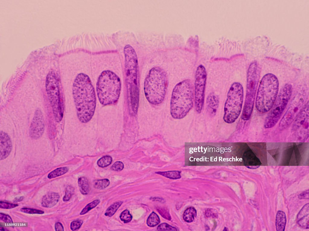
Found primarily in the respiratory epithelium, particularly lining the trachea and portions of the bronchi, ciliated simple columnar epithelium forms a continuous, stratified yet seemingly unbroken layer of cells. Each cell is tall and columnar in shape, its apical surface densely studded with motile cilia—microscopic hair-like projections that beating in synchronized waves propel mucus and trapped particles upward toward the throat. This rhythmic coordinated motion, often described as “the mucociliary escalator,” ensures continuous clearance of inhaled pollutants, bacteria, and allergens.
What makes this tissue exceptional is its dual role: structural integrity and functional agility. Each specialized cell contributes to a meticulously orchestrated defense system.
The nuclear membrane sits just beneath a large, centrally located nucleus, supporting the high metabolic demands of ciliary motion. Beneath it, the cell body houses abundant mitochondria and a well-developed endoplasmic reticulum, fueling ATP production essential for ciliary beating — a process requiring precise energy regulation. The most defining feature, however, is the apical mucous blanket and associated cilia.
Structure and Cellular Dynamics Explained
- **Ciliated Surface:** Columnar cells boast 200–300 cilia per cell, embedded in a gel-like periciliary liquid layer. This layer thins the surface for cilia to sweep efficiently across the epithelium, while maintaining optimal hydration to protect delicate ciliary structures. - **Mucus Layer:** Goblet cells interspersed among the columnar lining secrete a viscous mucus rich in glycoproteins and immunoglobulins. This sticky substance traps airborne particles, microbes, and debris, forming a protective barrier. - **Ciliary Coordination:** The beating pattern follows a directional waveform — all cilia strike simultaneously in a metachronal rhythm, creating a forward propulsion force that moves mucus one cell length per 6–8 seconds.
This steady, non-arrhythmic motion is critical for continuous airway cleaning.
- **Cell Turnover and Regeneration:** The overlying epithelium undergoes rapid renewal; stem cells in the basal layer continuously regenerate mature ciliated cells, typically replacing them every 48–72 hours. This turnover ensures rapid repair after injury or irritation. Beyond routine mucus clearance, this epithelium exhibits active defense mechanisms. Tight junctions between adjacent cells form a robust barrier, limiting pathogen penetration.
Additionally, epithelial cells express pattern recognition receptors that detect microbial components, triggering localized immune responses. Studies confirm that disruptions in ciglia or mucus quality — as seen in conditions like primary ciliary dyskinesia — significantly impair clearance, increasing susceptibility to chronic infections.
Clinical and Environmental Implications
The effectiveness of ciliated simple columnar epithelium directly impacts respiratory health.Environmental toxins, cigarette smoke, and chronic inflammation impair ciliary function and disrupt mucus composition. Smoking, for example, paralyzes cilia within minutes and destroys goblet cells, leading to impaired mucus transport and recurrent infections. Occupational exposures to dust, chemicals, and pollutants similarly compromise epithelial integrity, contributing to conditions such as chronic bronchitis or bronchiectasis.
In certain diseases, this epithelium undergoes metaplastic or dysplastic changes. Chronic irritant exposure can shift cell types — such as squamous metaplasia replacing ciliated columnar cells — weakening mucociliary clearance and predisposing to infection. Advances in pulmonary medicine now focus on regenerative strategies to restore epithelial function, including stem cell therapies and targeted modulation of ciliary beat regulators.
Ciliated simple columnar epithelium may operate unseen, but its absence would cripple the body’s frontline defense in the respiratory system. Through its seamless integration of structure, movement, and molecular signaling, this tissue exemplifies biological precision, ensuring that every breath remains clear and protected. Understanding its biology not only illuminates core physiological mechanisms but also guides therapeutic innovations for millions suffering from chronic respiratory disorders.


Related Post

Minecraft Alex: The Minecraft Wizard Making Code and Creativity Converge

Belkys Nerey Net Worth: The Rise of a Rising Star in Colombian Entertainment and Entrepreneurship

Understanding Sixy Video: Decoding the Next Wave of Digital Entertainment Transformation


