Decoding the Body: How the Transverse Anatomical Plane Revolutionizes Modern Medicine
Decoding the Body: How the Transverse Anatomical Plane Revolutionizes Modern Medicine
The human body is a three-dimensional marvel—complex, dynamic, and structured in ways that defy flat representation. Yet medical science relies on tools that translate this spatial reality into meaningful, actionable data. Among the most foundational of these tools is the transverse anatomical plane, a dividing line that slices the body horizontally, enabling precise orientation, visualization, and understanding of anatomical relationships.
This plane cuts through the vertical axis of symmetry, forming cross-sectional views that have transformed how clinicians diagnose, operate, and teach human anatomy. By orienting professionals in a universally accepted frame of reference, the transverse plane bridges the gap between abstract knowledge and real-world application, making it an indispensable concept across fields from surgery to imaging.
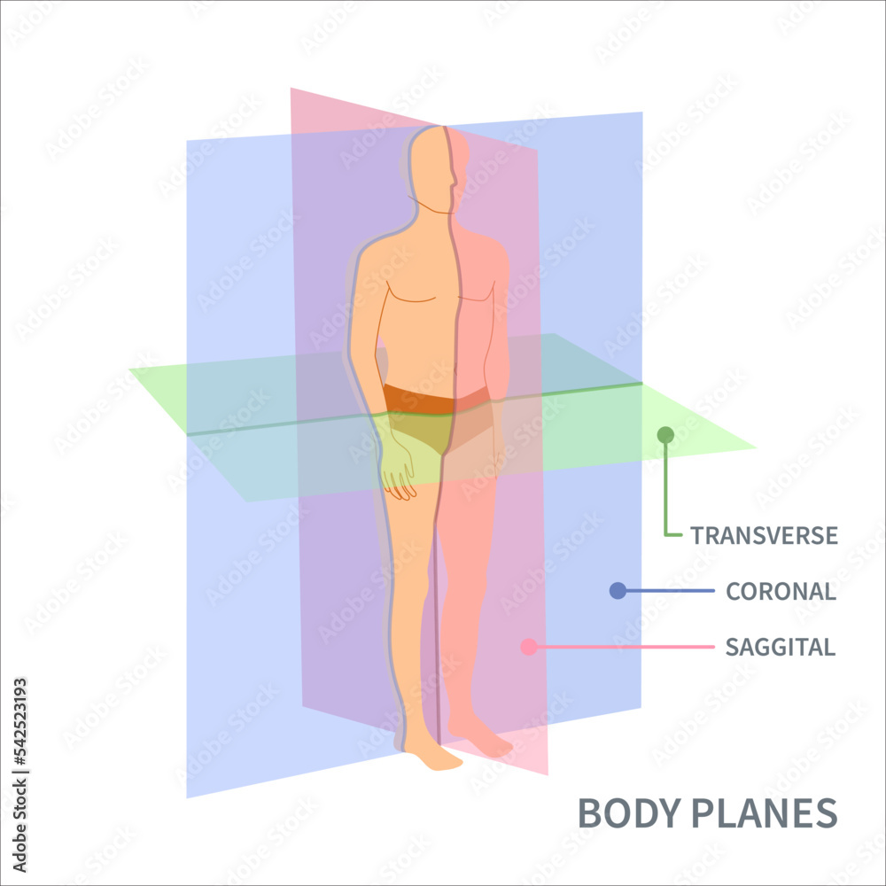
The transverse anatomical plane, also known as the transverse or axial plane, divides the body into superior (upper) and inferior (lower) segments.
Unlike the frontal (coronal) or sagittal planes—each offering distinct perspectives—the transverse plane provides a top-down, level view that mirrors natural body position. This alignment mirrors how surgeons approach procedures, how radiologists interpret scans, and how students mentally map anatomical structures. “The transverse plane is the gold standard for cross-sectional analysis,” notes Dr.
Elena Torres, a clinical anatomist at Stanford University. “It allows us to see organs, vessels, and tissues not as isolated entities but in functional relationships—exactly how they interact during disease or healing.”
One of the most significant strengths of the transverse plane lies in its compatibility with advanced imaging technologies. Modalities such as computed tomography (CT), magnetic resonance imaging (MRI), and ultrasound generate transverse slices that offer unprecedented clarity.
Each slice captures cross-sectional data with millimeter precision, enabling doctors to detect tumors, evaluate trauma, or monitor disease progression without invasive exploration. For instance, in emergency medicine, a CT scan using the transverse plane can rapidly reveal internal bleeding or organ lacerations, guiding life-saving interventions within minutes. “This plane transforms raw image data into diagnostic insight,” explains Dr.
James Cho, a radiologist at Mayo Clinic. “Without it, we’d be guessing—not seeing.”
Beyond diagnostics, the transverse plane serves as a cornerstone of surgical precision. During minimally invasive procedures, surgeons use real-time guidance based on transverse imaging to navigate delicate anatomy.
In robotic-assisted surgery, for example, the plane acts as a reference for tool placement, ensuring movements stay within safe, accurate corridors. For open procedures, it helps map tissue planes, identify critical landmarks, and assess resection margins—especially vital in oncological surgeries. “Every incision we make gains clarity from the transverse view,” says Dr.
Priya Mehta, an abdominal surgeon. “It’s not just imaging—it’s surgical intelligence.”
Educationally, the transverse plane has become the primary framework for teaching anatomy. Traditional cadaver labs integrate cross-sectional views derived from the transverse plane, helping students transition from two-dimensional diagrams to dynamic, spatial understanding.
Textbooks now feature annotated transverse slices, enabling readers to visualize organs in situ. Digital learning platforms take this further, offering interactive models where users can explore layer by layer. As Dr.
Torres observes, “Students who master the transverse plane develop a spatial literacy that enhances every clinical skill they’ll use.” Soaring demand for imaging proficiency in medical training underscores the plane’s educational dominance.
Clinically, the transverse plane supports targeted interventions across specialties. In interventional radiology, it enables precise needle placement for biopsies or drain insertions, minimizing risk to surrounding tissue.
In oncology, it aids in radiation therapy planning by defining tumor volumes and avoiding healthy tissue. Even in anesthesia, skeletal anatomy visualized in transverse sections guides nerve block placement. “It’s not just about seeing—it’s about guiding action,” says Dr.
Cho. “The plane ensures every treatment is anchored in anatomy, not assumption.”
The transverse anatomical plane has cemented its place as a linchpin of medical science. By offering a stable, standardized reference for imaging, surgery, education, and diagnosis, it transforms abstract human form into actionable knowledge.
Its influence spans from textbook diagrams to emergency rooms, from student classrooms to operating theaters. As technology advances—integrating AI with 3D reconstructions and augmented reality—the transverse plane remains the enduring foundation, ensuring clarity and precision in an increasingly complex field. This plane is not merely a section; it is the gateway to understanding the body’s inner architecture, one level at a time.


Related Post

Skyscraping Green: Unlocking Indonesia’s Vast Renewable Energy Potential
Shawn Spears Mocks Cody Rhodes His Unprotected Chair Shot