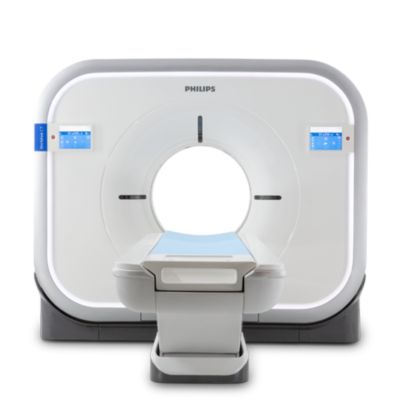
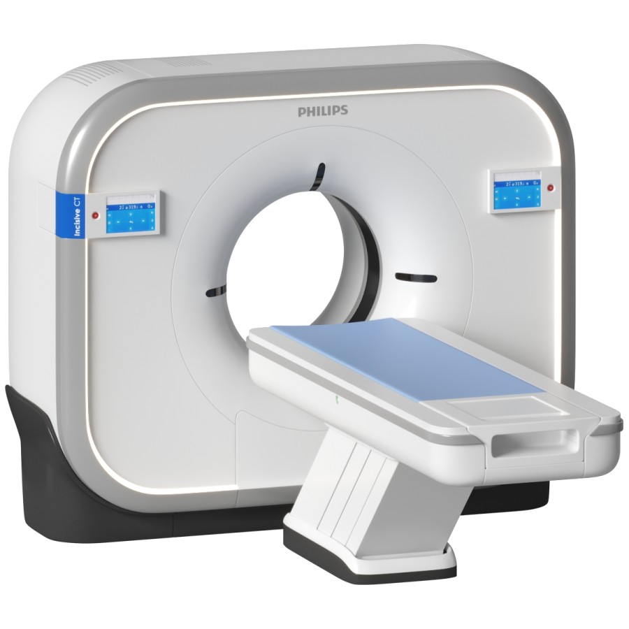
Philips Incisive CT: Your Go-To User Manual
Philips Incisive CT stands as a benchmark in modern medical imaging, combining speed, precision, and patient comfort into a single, powerful diagnostic tool. Designed for both urgent clinical scenarios and meticulous diagnostic workflows, the Incisive CT platform empowers radiologists and technologists with intuitive controls, advanced imaging algorithms, and real-time optimization—making it the trusted choice in hospitals and imaging centers worldwide. This comprehensive guide strips away complexity to present the essential insights every user needs to harness the full potential of Philips Incisive CT, from setup to scan execution and beyond.
Engineered Precision: The Core Capabilities of Philips Incisive CT
At the heart of Philips Incisive CT lies a robust architecture engineered for clinical excellence.The system delivers exceptional image quality with minimal radiation dose, making it ideal for repeated scans and sensitive patient populations. Renowned for its high-resolution imaging, Incisive CT achieves sub-millimeter spatial detail, enabling radiologists to detect subtle pathologies—such as early-stage tumors, micro-bleeds, and vascular abnormalities—with remarkable clarity. A standout feature is its intelligent scan engine, which dynamically adjusts acquisition parameters in real time.
This adaptability ensures optimal contrast resolution in challenging anatomies—whether imaging the lungs, abdomen, or neurovascular structures—without manual intervention. Clinicians report that this “smart” aspect reduces scan variability and improves consistency across high-volume departments. Advanced Detectors and Spectral Imaging form another cornerstone.
The dual-source technology, coupled with photon-counting-ready detectors, supports rapid, ultra-high-resolution acquisitions. Combined with spectral imaging capabilities, Incisive CT enables tissue characterization beyond conventional CT: differentiating iodine from calcium, enhancing lesion delineation, and supporting functional imaging workflows. These features are pivotal in oncology, cardiovascular, and pulmonary diagnostics, where accurate tissue classification directly impacts diagnosis and treatment planning.
Optimized Workflow and User Experience
Philips Incisive CT is built around the principle of seamless integration into fast-paced clinical environments. The user interface, developed with input from frontline technologists, prioritizes intuitive navigation—shortcuts, custom diagnostic templates, and customizable worklists reduce setup time and minimize cognitive load. Whether preparing for a trauma scan or a routine follow-up, staff can execute scans efficiently with confidence.Speed Meets Accuracy is the system’s defining performance trait. With rotational speeds up to 0.25 seconds per full rotation and helical pitch adjustments, spiral acquisition times are minimized—dropping silent scanning from seconds to under 10 seconds for key studies. This speed is critical in emergency departments and intensive care units, where rapid diagnosis can be life-saving.
Radiation Dose Management is equally refined. The Inscape Dose module leverages patient-specific algorithms and real-time dose modulation, maintaining diagnostic image quality while reducing radiation exposure by up to 50% compared to prior-generation systems. This aligns with evolving patient safety guidelines and regulatory demands, reinforcing Incisive CT’s role as a responsible choice in modern radiology.
Ergonomic Design and Patient Comfort further distinguish the platform. Patient islands feature adjustable tables, ambient lighting, and quiet scanning protocols—all designed to reduce anxiety, especially in pediatric, geriatric, and claustrophobic patients. Workflow-boosting features such as automatic table positioning and rapid access panels ensure comfort without sacrificing technical performance.
Clinical Applications: Where Incisive CT Delivers Impact
The versatility of Philips Incisive CT manifests across a broad spectrum of diagnostic applications. In neuroimaging, its speed and high contrast resolution enable undisturbed stroke assessments—critical for timely thrombolysis decisions. In cardiology, coronary CT angiography benefits from ultra-fast gantry rotation and motion correction, producing clear, motion-free images of coronary arteries, even in arrhythmic patients.Abdominal and pelvic imaging leverage the system’s superior spatial detail and spectral capabilities, allowing precise staging of oncology cases and characterization of complex liver lesions. For musculoskeletal applications, fast scanning enables high-quality evaluation of trauma patients, while advanced post-processing tools support 3D reconstruction and virtual endoscopy. Pediatric imaging exemplifies the system’s patient-centric engineering: low-dose protocols, child-friendly interfaces, and rapid acquisition keep children still and reduce sedation needs—an essential advantage in vulnerable populations.
Technical Mastery: From Setup to Post-Processing
Operational simplicity belies the platform’s technical depth. The boot-up sequence is streamlined for quick readiness, with automatic quality checks ensuring detector alignment and beam calibration prior to scan start. Technologists navigate through a unified software interface—accessible via touchpads or remote control—where protocols are preloaded with evidence-based settings for common exams, reducing setup errors.Real-time monitoring tools provide critical feedback during acquisition: radiation dose indicators, patient coupling sensors, and anatomical verification prompts help maintain quality and safety without disrupting workflow. Post-scan, integrated reconstruction software enables rapid processing—volume rendering, MPR, and 3D models generated in minutes, accelerating radiologist review. Advanced post-processing extends diagnostic reach: iodine maps highlight vascular pathology, lung windows enhance nodule detection, and motion correction tools stabilize images from breathing or cardiac motion.
These capabilities transform raw data into clinically actionable insights, supporting multidisciplinary care teams with precision.
Support, Updates, and Long-Term Value
Philips addresses the full lifecycle of clinical use with comprehensive support. Onsite service, remote diagnostics, and cloud-connected maintenance ensure minimal downtime—critical for 24/7 imaging centers.Regular software updates deliver performance enhancements, new protocols, and compliance enhancements without interrupting service. Training resources are intuitive and accessible: video tutorials, interactive modules, and peer networks help teams master advanced features efficiently. This commitment to continuous learning ensures adoption scales with technological evolution.
Cost-effectiveness is embedded in the platform’s design. Durable hardware, low maintenance demands, and energy-efficient operation reduce total cost of ownership. High scanner uptime and repeat clinical acceptance justify long-term investment, aligning with hospital budgets and patient care priorities.
Final Thoughts: Philips Incisive CT as the Imaging Benchmark
Philips Incisive CT is more than a scanner—it is a diagnostic partner built for the demands of modern medicine. Its fusion of speed, image fidelity, patient comfort, and intelligent workflow optimization positions it as the go-to choice for institutions committed to excellence in imaging. From urgent stroke assessments to detailed oncologic staging, the system delivers consistent, high-quality results with minimal operational friction.As clinical needs evolve, Incisive CT’s adaptable architecture and ongoing innovation ensure it remains a cornerstone of precision diagnostics.


Related Post

The Unbreakable Fire: The Band Members of Oasis and Their Lasting Musical Legacy
The Omega Ruby, Alpha Sapphire, and Pokémon OmegaRuby AlphaSapphire Pokedex: Unlocking the Ultimate Jungle Defense

Does Oshi No Ko Feature High-Octane Fight Scenes That Drive the Action?


