The Lateral Sulcus: Nature’s Clear Boundary Between Brain Lobes
The Lateral Sulcus: Nature’s Clear Boundary Between Brain Lobes
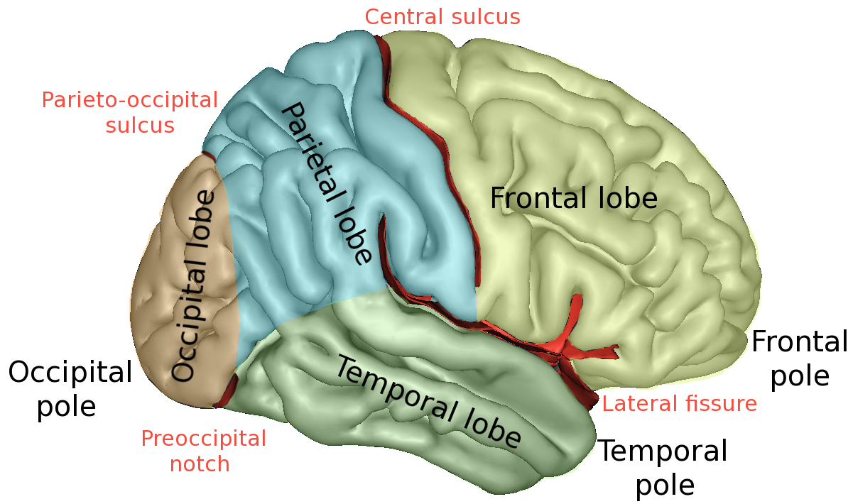
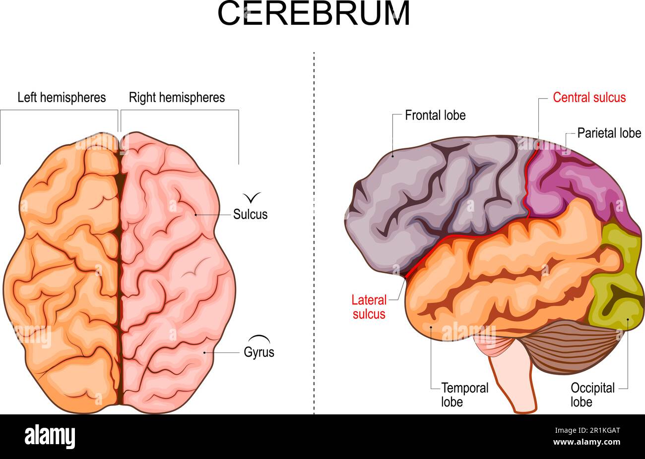
Deeper than a winding river cutting through a canyon, the lateral sulcus—the brain’s most prominent fold—serves as a precise anatomical and functional divider separating the frontal, parietal, temporal, and occipital lobes. This deep groove, running horizontally along the inferior edge of the cerebral hemisphere, is far more than a surface ridge: it is a masterful demarcation that guides both brain organization and clinical understanding. For neuroscientists and clinicians alike, the lateral sulcus defines where one lobe begins and another ends, shaping how sensory input, motor control, and higher cognition are processed across the brain’s landscape.
Anatomical Landmark: The Shape and Significance of the Lateral Sulcus
The lateral sulcus, also known as Sylvius’s sulcus after the 17th-century anatomistFounder of its detailed description, stretches from the rear of the frontal cortex near the parietal-occipital junction to the front of the temporal lobe where it meets the parieto-occipital sulcus. It appears as a sharp, arched furrow, varying subtly in depth and location across individuals—a testament to the brain’s unique topography. Despite its irregular contour, its function remains consistent: a hard-wired boundary that isolates distinct cortical territories.This sulcus arises just behind the precuneus, a critical region involved in self-awareness and memory, and descends nearly horizontally toward the zlf silk 1 and temporal pole. Its position creates a natural segregation: superior regions give way to temporal and parietal lobes below, while frontal functional zones remain distinctly above. This anatomical precision enables efficient neural circuitry, ensuring signals remain confined within their intended lobe domains—critical for accurate perception, reasoning, and motor coordination.
Separating Lobes: Which Structures Lie on Each Side
To the lateral (outer) side of the lateral sulcus—encompassing the region running from near the parietal lobe’s tentorium to the temporal pole—lie key lobes: frontal, parietal, temporal, and part of the occipital lobe. The frontal lobe, responsible for executive function, decision-making, and voluntary movement, begins just dorsal and superior to the sulcus. Moving laterally, the parietal lobe—integral to spatial awareness and sensory integration—occupies the space to the lateral sulcus’s core.Below this ridge, the temporal lobe dominates, housing auditory processing, memory consolidation, and language comprehension. Even the occipital lobe, primarily visual, extends slightly lateral here, though much of its bulk lies posteriorly. Below the lateral sulcus’s deepest point, the insular cortex dips in—a fold hidden but vital, separating temporal functionality from the deeper insula beneath the Sylvian fissure.
The sulcus thus neatly isolates: - **Frontal lobe**: Control, planning, and motor command - **Parietal lobe**: Sensory mapping and spatial navigation - **Temporal lobe**: Auditory, memory, and language centers - **Insular cortex**: Interoception and emotional processing (via its lower portion) Each lobe functions with a degree of autonomy, enabled by this clear anatomical partitioning.
Functional Specialization Across the Sulcus’s Divide
While the lateral sulcus serves as a structural divider, its influence extends into the realm of specialized brain functions. Across its divide lies a functional zonation that underpins human cognition.The frontal lobe, with its robust prefrontal cortex, governs impulse control, strategic thinking, and goal-directed behavior. Damage here—whether from stroke or trauma—often disrupts executive function, revealing just how vital this lobe’s unbroken territory is. Across to the parietal lobe, spatial reasoning flourishes.
Studies using fMRI consistently activate this region during navigation, manipulation of objects, or mental rotation tasks. The precise boundary created by the sulcus ensures that sensory input from both hemispheres converges meaningfully but remains contextually segregated—allowing spatial awareness to operate without interference from motor planning or language centers nestled dorsally. The temporal lobe, shielded subtly by the sulcus, becomes the epicenter of memory encoding and auditory discrimination.
The lateral affiliation here supports the hippocampus’s role in forming episodic memories and Wernicke’s area’s critical part in language comprehension—functions so lateralized that impaired integration could severely fragment communication and recall. Even the insular cortex, partially tucked beneath the sulcus, benefits from its cleavage: it mediates emotional awareness and bodily sensation, functions that interface with both limbic (temporal) and prefrontal (frontal) networks. This topological isolation enables nuanced emotional regulation, seamlessly blending internal states with cognitive appraisal.
Clinical Implications: When the Sulcus Marks Dysfunction
Understanding the lateral sulcus’s role is not merely academic—it directly informs neurological diagnosis and treatment. Imaging modalities like MRI rely on its consistent anatomical landmarks to detect abnormalities. Enlargement or deepening of the sulcus, for instance, can signal cortical atrophy seen in degenerative diseases such as Alzheimer’s, where temporal lobe shrinkage disrupts memory systems.Similarly, stroke affecting mid-sylvian regions often impairs language (Broca’s or Wernicke’s areas) or spatial processing, depending on the exact sulcus-adjacent lobe injured. Surgical planning also hinges on mapping the lateral sulcus with precision. Neurosurgeons navigating brain tumors or epilepsy must avoid breaching this sulcus canyons to prevent dismantling critical functional areas.
The sulcus acts as a natural guide, delineating safe zones where resection is possible without catastrophic lobe damage. Beyond pathology, the lateral sulcus informs our understanding of brain plasticity. As rehabilitation progresses after injury, shifts in sulcal proximity—driven by reorganizing neural networks—can reflect functional recovery, offering clinicians quantifiable markers of healing.
In research, the lateral sulcus remains a focal point for uncovering how cerebral architecture supports the mind. Advanced neuroimaging continues to refine its boundary definitions, revealing subtle variations linked to individual cognitive profiles, learning styles, and even artistic or linguistic aptitude. This evolving map underscores the sulcus not as a static line, but as a dynamic interface shaped by both genetics and experience.
The Lateral Sulcus: A Window into Brain Architecture and Mind
The lateral sulcus is far more than a groove etched into the brain’s surface—it is a master classifier, a precise anatomical sentinel that carves the cerebral landscape into functionally distinct lobes. By separating the frontal, parietal, temporal, and insular regions, it enables the specialized processing that defines human cognition. From guiding motor commands to orchestrating memory and language, each lobe’s activity thrives within the boundaries defined by this relentless fold.As neuroscience advances, the lateral sulcus remains central to both curiosity and clinical practice—reminding us that even the deepest sulci hold the clearest keys to understanding the mind. This enduring anatomical feature, sculpted by evolution and refined by biology, ensures that the brain’s complexity unfolds not in chaos, but in exquisite, biologically meaningful order.


Related Post

How P.O. Boxs Are Transforming Secure Mail Delivery in the Digital Age

Oso Panda: Nature’s Master of Camouflage – Unveiling Its Truly Unique Superpowers

Scam Or Legit? Decoding the Truth Behind IBullMarket.com’s Emerging Reputation

Watch Galveston’s Port Live: Real-Time Cruise Cam Offers a Peek into the Heart of Texas’s Coastal Gateway