Vertical Angulation Dental Radiography: The Precision Tool Shaping Modern Dentistry
Vertical Angulation Dental Radiography: The Precision Tool Shaping Modern Dentistry
In the ever-evolving landscape of dental diagnostics, vertical angulation radiography stands as a cornerstone imaging technique, delivering unparalleled accuracy in visualizing tooth anatomy and pathology. Unlike conventional radiographs, vertical angulation—often referred to as vertical True Field Imaging (VTFI)—precision measures the true vertical dimension of dental structures, ensuring that measurements are not distorted by angulation errors. This advancement has transformed how clinicians approach endodontic treatment planning, implant placement, and assessment of periodontal health.
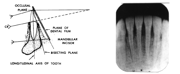
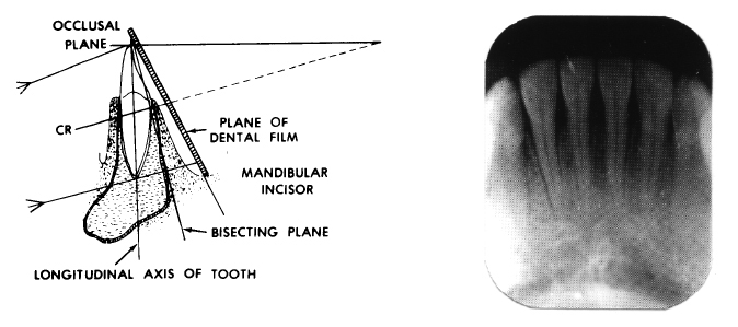
By capturing animage aligned with the actual vertical axis of teeth, this technique delivers diagnostic clarity that enhances both detection and intervention. Understanding vertical angulation begins with its core principle: aligning the X-ray source and detector perpendicular to the tooth’s long axis. Traditional radiographic images often suffer from geometric distortion when angled incorrectly, leading to misjudgments in root length, bone levels, and lesion depth.
Vertical angulation radiography eliminates this pitfall by maintaining a perpendicular orientation, resulting in radiographs that faithfully represent the true three-dimensional relationships within the oral cavity. As Dr. Elena Marquez, a leading oral radiologist, explains, “Vertical angulation ensures that measurements reflect real anatomy—not the distortion caused by camera tilt.” This precision is particularly vital in identifying vertical bone defects, assessing root resorption, and verifying proper apexification in vital pulp therapy.
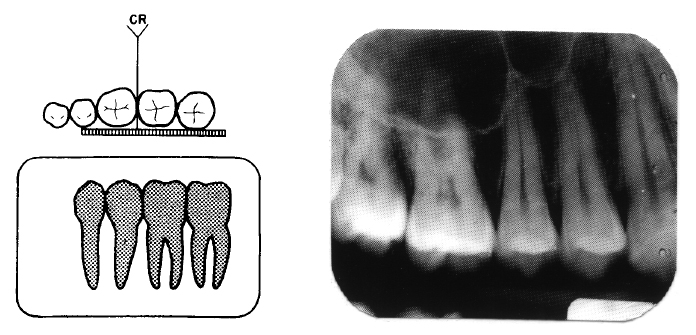
At the technical core of vertical angulation radiography is the alignment of imaging components within a specific geometric matrix. The X-ray beam is emitted directly perpendicular to the tooth surface, typically using a vertical cone beam or intraoral digital sensor positioned parallel to the occlusal plane. This configuration preserves the true vertical dimension, enabling unambiguous visualization of critical structures such as the alveolar crest, neural foramina, and pulp chambers.
Unlike enfilade imaging, which captures teeth in a diagonal plane and introduces depth inaccuracies, vertical angulation delivers a true-mile diagnostic window.
- Vertical Angulation vs. Conventional Radiography: Traditional techniques often amplify measurement variance due to camera positioning deviations, particularly in posterior teeth with complex root anatomy. Studies show vertical angulation reduces vertical error by up to 60%, significantly improving diagnostic reliability.
- Digital Integration: Most modern systems combine vertical angulation principles with digital radiography, offering real-time image preview, dynamic vertical calibration, and reduced radiation exposure.
This integration enhances workflow efficiency and patient safety.
- Clinical Applications: dentists use vertical angulation imaging routinely in endodontic diagnosis—mapping root canal morphology and detecting periapical lesions—and in implant dentistry, where accurate bone height and symmetry are paramount. It also excels in diagnosing vertical bone loss around resorbing teeth.
The benefits of vertical angulation extend beyond pure diagnostics into treatment execution. For endodontists, precise apical transverse measurements guide root canal instrumentation and filling depth.
Orthodontists rely on accurate root angulation data to plan extractions and evaluate tooth movements. In periodontics, vertical radiographs reveal subtle crestal bone defects invisible on standard bitewings, enabling early intervention. “Every millimeter matters in implant dentistry,” notes Dr.
Amir Patel, a clinical specialist. “Any miscalculation in vertical dimension could compromise osseointegration and long-term success.”
- Accessibility and Training
- While the technology enhances accuracy, adoption depends on proper training and equipment calibration. Dental schools now integrate VTFI modules into simulation labs to ensure practitioners master vertical angulation early in education.
Calibration of sensors and software algorithms remains essential to maintain geometric fidelity across devices.
- Radiation Efficiency
- Despite extended exposure windows compared to 2D films, vertical imaging systems often deliver doses within safe limits due to localized targeting. This precision reduces patient exposure without sacrificing diagnostic power—critical in preventive and routine care.
Current innovations continue refining vertical angulation’s utility. Emerging software now employs AI-assisted analysis to highlight vertical defects automatically, reducing interpretation time and human error.
Additionally, ergonomic designs of portable vertical X-ray units are expanding access in community clinics and teledentistry platforms, democratizing high-accuracy imaging. Beyond technical specs, vertical angulation radiography represents a paradigm shift toward measurement integrity in dentistry. By anchoring all diagnostic data to true vertical anatomy, it elevates care from reactive to predictive.
“This is not just about better images—it’s about better outcomes,” asserts Dr. Lisa Chen, a proponent of digital precision dentistry. As this technology becomes standard, dental professionals are empowered with the tools to deliver more accurate, personalized, and reliable treatment.
In the broader context of digital dentistry, vertical angulation radiography stands out as a critical enabler—ensuring every diagnostic step aligns with anatomical truth. As lasers, AI, and 3D imaging advance, this foundational geometry remains indispensable. It transforms radiographs from static images into dynamic diagnostic resources, shaping the future of oral healthcare with every precisely angulated exposure.
Vertical angulation dental radiography is not merely a technical detail—it is the backbone of precision in modern dental practice, ensuring that every treatment decision rests on accurate vertical anatomy.

Related Post
What Is Vertical Angulation Dental Radiography?
Fan Video Captures Sasha Banks Overcome With Happiness After WrestleMania Loss
Arizona Robbins: Architect of Grey Sloan Academy and Pioneer of Forensic Medicine


